The Brandberg–Roberts–Stolnikov method refers to semi-quantitative methods for determining total protein in urine. The method is based on Heller's ring test, which consists in the fact that at the border of nitric acid and urine, in the presence of protein, it coagulates and a white ring appears.
Reagents
50% nitric acid solution or Larionova's reagent.
Preparation of Larionova's reagent: prepare a saturated solution of sodium chloride (20 - 30 g of salt are dissolved in 100 ml of water when heated, let stand until cooled). The supernatant liquid is drained and filtered. To 99 ml of filtrate add 1 ml of concentrated nitric acid. Instead of nitric acid, you can add 2 ml of concentrated hydrochloric acid.
Progress of determination
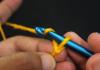
1 - 2 ml of nitric acid (or Larionic reagent) is poured into the test tube, the acid is allowed to drain from the walls of the test tube (5 - 8 minutes), otherwise, when protein urine is layered, turbidity will form due to the mixing of nitric acid on the walls of the test tube with urine, which prevents the formation of a distinct ring . Therefore, you should first prepare a series of test tubes with acid. Using a pipette, carefully layer the same amount of filtered liquid along the wall of the test tube. clear urine, being careful not to agitate the liquid in the test tube. The appearance of a thin white ring at the interface of the two liquids between the 2nd and 3rd minutes indicates the presence of protein at a concentration of approximately 0.033 g/l. Layering time is calculated as a quarter of a minute.
If the ring appears earlier than 2 minutes after layering, the urine should be diluted with water and the already diluted urine should be layered again. The degree of urine dilution is selected depending on the type of ring, i.e. its width, compactness and time of appearance. If a thread-like ring appears before 2 minutes, the urine is diluted 2 times, if it is wide - 4 times, if it is compact - 8 times, etc. Dilution of urine is done in a measuring centrifuge tube, pouring urine to the 1 ml mark and adding water to the mark by how many times the dilution is made. The contents of the test tube are thoroughly mixed with a Pasteur pipette and a balloon. If turbidity appears when diluting urine, the mixture must be filtered again and only the clear filtrate should be layered with nitric acid. The protein concentration is calculated by multiplying 0.033 by the degree of dilution and expressed in grams per liter (g/l). Select a dilution of urine such that when it is layered on nitric acid, a ring appears in the 2nd – 3rd minute.
If a ring forms with undiluted or diluted urine between the 1st and 4th minute, you can use the Ehrlich-Althausen correction in order not to dilute the urine additionally (this saves time). The authors proposed to determine the time of appearance of the thread-like ring and introduce a time correction into the calculation. In this case, the amount of protein is calculated by multiplying 0.033 g/l by the degree of dilution and the correction.
When a ring forms, before 1 minute has elapsed, it is necessary to make one fractional dilution, namely 1.5 times (two parts urine and 1 part water). This dilution is also taken into account when calculating the amount of protein in the urine.
An example of determining total protein in urine using the Brandberg-Roberts-Stolnikov method.
When urine is layered onto the reagent, a wide ring immediately forms. Dilute urine 4 times (1 part urine + 3 parts water), layer; A thread-like ring is immediately obtained. It is necessary to dilute the existing dilution by another 2 times; When layering this dilution, a ring is formed after 1.5 minutes. You don't have to breed any further.
Protein calculation: urine was diluted 4 and 2 times, therefore 8 times. The amount of protein is 0.033*8*1 1/4 = 0.33 g/l
Disadvantages of the Brandberg-Roberts-Stolnikov method:
- subjectivity,
- labor intensity,
- decrease in the accuracy of determining protein concentration as urine is diluted.
See also:
Literature:
- Handbook "Laboratory research methods in the clinic" ed. prof. V.V. Menshikova. - Moscow, "Medicine", 1987
- L. V. Kozlovskaya, A. Yu. Nikolaev. Tutorial on clinical laboratory research methods. Moscow, Medicine, 1985
- A. Ya. Althauzen, “Clinical laboratory diagnostics” - Moscow, Medgiz, 1959
- A. Ya. Lyubina, L. P. Ilyicheva and co-authors, “Clinical laboratory studies”, Moscow, “Medicine”, 1984
Solutions: 50% nitrogen solution or larion solution. Determination procedure: a row of test tubes is placed in a stand and 1 ml of nitrogen solution is poured, add 1 ml of urine, layered on the reagent and the time is noted; when a ring appears, we record the time of ring appearance. If the ring is wide, dilute the urine.
4. Determination of protein concentration in urine with 3% sulfosalicylic acid.
Solutions: 3% CK, sodium chloride 9%, albumin solution 10%. Determination procedure: 1.25 ml of clear urine is placed in two measuring centrifuge tubes “O” - experiment and “K” - control. Add 3.75 ml of 3% sulfosalicylic acid solution to the experimental one, and 3.75 ml of 0.9% sodium chloride solution to the control one. Leave for 5 minutes, and then photometerize on FEC at a wavelength of 590 - 650 nm (orange or red filter) in a cuvette with a layer thickness of 5 mm, experiment versus control. The calculation is carried out according to the calibration schedule or table. Principle of the method is based on the fact that protein with sulfosalicylic acid produces turbidity, the intensity of which is directly proportional to the protein concentration.
5. detection of glucose in urine Gaines-Akimov test. Principle: Glucose, when heated in an alkaline environment, reduces copper dihydroxide ( yellow color) to copper monohydroxide (orange-red color). Preparation of the reagent: 1) 13.3 g chemical. pure crystalline copper sulfate (CuSO 4 . 5 H 2 O) solution. in 400 ml of water. 2) 50g of sodium hydroxide is dissolved in 400ml of water. 3) 15g of pure glycerin is diluted in 200ml of water. Mix the first and second solutions and immediately add the third. Reagent racks. Progress of determination: Add 1 drop of urine and 9 drops of reagent into a test tube and boil in a water bath for 1-2 minutes. Positive test: yellow or orange color of liquid or sediment.
6. Qualitative definition glucose in urine using the glucose oxidase method. Principle of the method: glucose is oxidized in the presence of glucose oxidase, according to the reaction: Glucose + O 2 glyconolantom + H 2 O 2. The resulting peroxide H under the action of peroxidase oxidizes the substrate to form a colored product.
Add and incubate for 15 minutes at 37 0 C. Look at the CPK, 5mm cuvette.
Then calculations are made using the formula: C op = Ext op . Cst/ Ect st.
7. Detection of ketone bodies in urine by Lestrade test. A powder or tablet of Lestrade solution is applied to a glass slide (at the tip of a scalpel), and 2-3 drops of urine are applied to it. If ketone bodies are present, a pink to purple color will appear. The sample is evaluated against a white background.
8. Detection of blood pigment in urine by testing with a 5% alcohol solution of amidopyrine.
1.5% alcohol solution amidopyrine (0.5 g amidopyrine dissolved in 10 ml of 96% alcohol) 2.3% peroxide solution hydrogen, 1.5 g of hydropyrite is dissolved in 50 ml of water) Procedure: 2-3 ml of acetic ether extract or shaken unfiltered urine is poured into a test tube. 8-10 drops of 5% amidopyrine solution and 8-10 drops of 3% are added solution of hydrogen peroxide; take into account the result no later than 2-3 minutes. The sample is considered positive if it has a gray-violet color.
Detection of urobilin in urine by Neubauer test.
It is based on the color reaction of urobilinogen with Ehrlich's reagent, which consists of 2 g of paradimethylaminobenaldehyde and 100 ml of hydrochloric acid solution (200 g l). Determination procedure: A few drops of Ehrlich's solution are added to several ml of freshly excreted urine (on 1 ml of urine and per 1 ml of solution. The appearance of a red color in the first 30 s indicates an increase in urobilinogen. Normally, the color appears later or is absent altogether. When urine stands, urobilinogen turns into urobilin and the sample may be false negative. Sample cannot be heated, as side complex compounds of aldehyde with porphyrins, indole and medications may form.
Detection of bilirubin in urine by Rosin's test.
Alcohol solution of iodine (10 g.l): 1 g of crystalline iodine is dissolved in a cylinder with a capacity of 100 ml in 20-30 ml of 96 g of rectified alcohol, and then added with alcohol to the mark. Determination procedure. Pour into a chemical test tube 4-5 ml of the test urine and carefully layer an alcohol solution of iodine on it (if the urine has a low relative density, then it should be layered on an alcohol solution of iodine). If there is bilirubin at the boundary between the liquids, there will be a green ring (when taken antipyrine, as well as when there is soda in the urine, the blood pigment test turns out to be positive). In a healthy person, this test is negative.
Urine examination using dry chemistry method (mono-polytests).
Principle. The method is based on the effect that a protein has on the color of an indicator in a buffer solution, as a result of which the dye changes color from yellow to blue.
When carrying out a reaction for the presence of protein in urine and determining the pH using indicator paper, it is recommended to follow the following instructions:
- Collect urine in thoroughly washed dishes.
- Use freshly collected, preservative-free urine.
- Close the pencil case carefully after removing it from it. required quantity indicator strips of paper.
- Do not touch the indicator zones with your fingers.
- Use only within the expiration date indicated on the label.
- Follow the rules for storing indicator paper.
- Evaluate the results in accordance with the instructions in the instructions.
Performing a urine test using a urine dry chemistry analyzer.
Progress of determination. A strip of indicator paper is removed from the pencil case and immersed in the urine being tested so as to simultaneously wet both indicator zones. After 2-3 seconds, the strip is placed on a white glass plate. Immediately assess the pH using the color scale on the pencil case. The pH value on the color scale corresponds to 6.0 (or less); 7.0; 8.0; 9.0.
Preparation of urine, preparation of preparations from urine sediment for microscopic examination in an approximate manner.
Microscopic examination of urine sediment is carried out using the approximate method when general analysis and quantitative counting of formed elements for a more accurate assessment of the degree of luikocyturia and hematuria.
Rules for preparing urine sediment for microscopy.
The first morning portion of urine is subject to microscopic examination.
After preliminary mixing, 10 ml of urine is taken and centrifuged for 10 minutes at 1500 rpm.
Then the centrifuge tube with urine is overturned with a sharp movement, and the supernatant is quickly poured into an empty jar.
Mix, place a drop on a glass slide and carefully cover with a coverslip.
If the sediment consists of several layers, then prepare a preparation, and then centrifuge again and prepare preparations from each layer separately.
If there is no sediment visible to the eye, a drop of urine is applied to a glass slide and microscopically examined.
At the beginning, the material is examined at low magnification (eyepiece 7-10, objective 8), the condenser is lowered, the aperture is slightly narrowed, then the preparation is studied in detail at high magnification(eyepiece 10.7; objective 40).
14.Quantitative study of urine sediment according to Nechiporenko.
The method is used for latent, sluggish inflammatory processes (pyelonephritis, glomerulonephritis), latent pyuria. To study the pathological process in dynamics. To assess the effectiveness of the treatment. Advantages of the method: technically simple, does not require a large amount of urine and is long-lasting. its storage is used in outpatient practice. Obligation conditions: morning urine, medium portion, acidic solution (in alkaline urine there may be partial disintegration of cellular elements). 1. Mix the urine. 2. Place 10 ml of urine into a measuring centrifuge tube and centrifuge for 10 minutes at 1500 rpm. 3. After centrifugation. sucking Pipette the top of the liquid, leaving. exactly 1 ml of sediment. 4. The sediment is thoroughly mixed and Goryaev’s chamber is filled. 5. 3-5 minutes after filling, begin counting the formed elements. 6. Counting leukocytes, Er, cylinders with eyepiece 15, lens 8 with omission. condenser, in 100 large square chambers. Leukocytes, Er are counted separately, cylinders (at least 4 Goryaev chambers are counted) and the medium is removed. arif. X=A x 0.25x 10 6 /l. Norm: leuk. 2-4x 10 6 /l, Er up to 1 x 10 6 /l, cylinders up to 0.02 x 10 6 /l (one for 4 chambers). In children: leukemia. up to 2-4x 10 6 /l, Er up to 0.75 x 10 6 /l, cylinders up to 0.02 x 10 6 /l.
15. Urine examination according to Zimnitsky
This test determines the ability of the kidneys to concentrate. and dilute the urine. Essence of sample seal. in dynamic definition relative density and the amount of urine in three-hour portions during the day. Conducting the test: after emptying Bladder at 6 o'clock in the morning in the toilet, the patient collects urine into separate jars every three hours during the day. Total 8 servings. Progress of the research: 1. Delivery. Urine is placed by the hour and the quantity and relative density are determined in each portion. 2. Compare the daily amount of urine and the amount of liquid drunk to determine. % of its excretion. 3. Calculate daytime and nighttime diuresis, sum it up, and get daily diuresis. 4. Set the range of fluctuations in quantity and relative. urine density per day i.e. what is the difference between the smallest portion and the largest. Show. samples from healthy people people: 1. Daily diuresis 800-1500 ml. 2. Daytime diuresis significantly prevails over nighttime diuresis. 3. Fluctuations in the volume of urine in individual portions are significant (from 50 to 400 ml). 4. Fluctuations p from 1.003 to 1.028, should be more than 0.008. With function renal failure: hyposthenuria, hypoisosthenuria, isosthenuria, hypersthenuria, oliguria, anuria, nocturia.
16. Description of the general properties of feces.
Normally, feces consists of secretion and excretion products of the digestive tract, undigested or partially digested food residues, and microbial flora. The amount of feces is 100-150 g. The consistency is dense. The shape is cylindrical. The smell is normal. Brown color. R-tion - neutral, slightly alkaline or slightly acidic (pH 6.5-7.0-7.5). Mucus-absent. Blood-absent. There are no remains of undigested food.
Protein in urine: methods of determination
Pathological proteinuria is one of the most important and permanent signs of kidney and urinary tract diseases. Determination of protein concentration in urine is a mandatory and important element of urine testing. Identification and quantitative assessment of proteinuria is important not only in the diagnosis of many primary and secondary kidney diseases; assessment of changes in the severity of proteinuria over time carries information about the course of the pathological process and the effectiveness of the treatment. Detection of protein in the urine, even in trace amounts, should raise red flags for possible kidney or urinary tract disease and require repeat testing. Particularly noteworthy is the pointlessness of urine examination and, in particular, determination of urine protein without compliance with all rules for its collection.
All methods for determining protein in urine can be divided into:
High quality,
Semi-quantitative,
Quantitative.
Qualitative methods
All qualitative tests for protein in urine based on the ability of proteins to denature under the influence of various physical and chemical factors. If protein is present in the urine sample being tested, either turbidity or flocculent sediment appears.
Conditions for determining protein in urine based on the coagulation reaction:
The urine should be acidic. Alkaline urine is acidified with a few (2 - 3) drops of acetic acid (5 - 10%).
Urine should be clear. Cloudiness is removed through a paper filter. If the turbidity does not disappear, add talc or burnt magnesia (about 1 teaspoon per 100 ml of urine), shake and filter.
A qualitative sample should be carried out in two test tubes, one of them is a control one.
You should look for haze on a black background in transmitted light.
Qualitative methods for determining protein in urine include:
Heller ring test,
test with 15 – 20% sulfosalicylic acid,
boiling test, and others.
As numerous studies show, none of the large number known methods for the qualitative determination of protein in urine do not allow obtaining reliable and reproducible results. Despite this, in most DDLs in Russia these methods are widely used as screening - in urine with a positive qualitative reaction, protein is subsequently quantitatively determined. Of the qualitative reactions, the Heller test and the test with sulfosalicylic acid are most often used, but the test with sulfosalicylic acid is generally considered the most suitable for identifying pathological proteinuria. The boiling test is currently practically not used due to its labor intensity and duration.
Semi-quantitative methods
TO semi-quantitative methods relate:
Brandberg-Roberts-Stolnikov method,
determination of protein in urine using diagnostic test strips.
The Brandberg-Roberts-Stolnikov method is based on the Heller ring test, so with this method the same errors are observed as with the Heller test.
Currently, diagnostic strips are increasingly used to determine protein in urine. For the semi-quantitative determination of protein in urine on a strip, the dye bromophenol blue in citrate buffer is most often used as an indicator. The protein content in urine is judged by the intensity of the blue-green color that develops after contact of the reaction zone with urine. The result is assessed visually or using urine analyzers. Despite the great popularity and obvious advantages of dry chemistry methods (simplicity, speed of analysis), these methods of urine analysis in general and protein determination in particular are not without serious drawbacks. One of them, leading to distortion of diagnostic information, is the greater sensitivity of the bromophenol blue indicator to albumin compared to other proteins. In this regard, test strips are mainly adapted to detect selective glomerular proteinuria, when almost all urine protein is albumin. With the progression of changes and the transition of selective glomerular proteinuria to non-selective (appearance of globulins in the urine), the results of protein determination turn out to be underestimated compared to the true values. This fact does not make it possible to use this method determination of protein in urine to assess the condition of the kidneys (glomerular filter) over time. With tubular proteinuria, the results of protein determination are also underestimated. Protein testing with test strips is not a reliable indicator of low levels of proteinuria (most currently available test strips are not capable of detecting urinary protein concentrations lower than 0.15 g/L). Negative results of protein determination on strips do not exclude the presence of globulins, hemoglobin, uromucoid, Bence Jones protein and other paraproteins in the urine.
Flakes of mucus with a high content of glycoproteins (for example, during inflammatory processes in the urinary tract, pyuria, bacteriuria) can settle on the indicator zone of the strip and lead to false-positive results. False positive results may also be due to high concentrations urea. Poor lighting and impaired color vision can cause inaccurate results.
In this regard, the use of diagnostic strips should be limited to screening procedures, and the results obtained with their help should be considered as indicative only.
Quantitative methods
Correct quantitative determination of protein in urine in some cases it turns out to be a difficult task. The difficulties of solving it are determined by the following number of factors:
the presence in the urine of many compounds that can interfere with the course of chemical reactions;
significant fluctuations in the content and composition of urine proteins during various diseases, making it difficult to select an adequate calibration material.
In clinical laboratories, so-called “routine” methods for determining protein in urine are mainly used, but they do not always provide satisfactory results.
From the point of view of a laboratory analyst, a method intended for the quantitative determination of protein in urine must meet the following requirements:
have a linear relationship between the absorption of the complex formed during the chemical reaction and the protein content in the sample over a wide range of concentrations, which will avoid additional operations when preparing the sample for research;
should be simple, not require high qualifications of the performer, and be performed with a small number of operations;
have high sensitivity and analytical reliability when using small volumes of test material;
be resistant to various factors (fluctuations in the composition of the sample, the presence of drugs, etc.);
have an acceptable cost;
be easily adaptable to auto analyzers;
the determination result should not depend on the protein composition of the urine sample being tested.
None of the currently known methods for the quantitative determination of protein in urine can fully claim to be the “gold standard”.
Quantitative methods for determining protein in urine can be divided into turbidimetric and colorimetric.
Turbidimetric methods
Turbidimetric methods include:
protein determination with sulfosalicylic acid (SSA),
protein determination with trichloroacetic acid (TCA),
determination of protein with benzethonium chloride.
Turbidimetric methods are based on a decrease in the solubility of urine proteins due to the formation of a suspension of suspended particles under the influence of precipitating agents. The protein content in the test sample is judged either by the intensity of light scattering, determined by the number of light-scattering particles (nephelometric method of analysis), or by the attenuation of the light flux by the resulting suspension (turbidimetric method of analysis).
The amount of light scattering in precipitation methods for detecting protein in urine depends on many factors: the rate of mixing of reagents, the temperature of the reaction mixture, the pH value of the medium, the presence of foreign compounds, and photometric methods. Careful adherence to reaction conditions will result in the formation of a stable suspension with a constant particle size and relatively reproducible results.
Some medications affect the results of turbidimetric methods for determining protein in urine, leading to so-called “false positive” or “false negative” results. These include some antibiotics (benzylpenicillin, cloxacillin, etc.), radiocontrast iodine-containing substances, and sulfonamide drugs.
Turbidimetric methods are difficult to standardize and often lead to erroneous results, but despite this, they are currently widely used in laboratories due to the low cost and availability of reagents. The most widely used method in Russia for protein determination is sulfosalicylic acid.
Colorimetric methods
The most sensitive and accurate are colorimetric methods for determining total urine protein, based on specific color reactions of proteins.
These include:
biuret reaction,
Lowry method,
methods based on the ability of various dyes to form complexes with proteins:
Ponceau S,
Coomassie Brilliant Blue
pyrogallol red.
From the point of view of the performer, in the daily work of the laboratory with a large flow of research, the biuret method is inconvenient due to the large number of operations. At the same time, the method is characterized by high analytical reliability, allows the determination of protein in a wide range of concentrations and the detection of albumin, globulins and paraproteins with comparable sensitivity, as a result of which the biuret method is considered as a reference and is recommended for comparison of other analytical methods for detecting protein in urine. The biuret method for determining protein in urine is preferably performed in laboratories serving nephrology departments, and is used in cases where the results of determination using other methods are questionable, as well as to determine the amount of daily protein loss in nephrology patients.
The Lowry method, which is more sensitive than the biuret method, combines the biuret reaction and the Folin reaction to the amino acids tyrosine and tryptophan in the protein molecule. Despite its high sensitivity, this method does not always provide reliable results when determining protein content in urine. The reason for this is the nonspecific interaction of Folin's reagent with non-protein components of urine (most often amino acids, uric acid, carbohydrates). The separation of these and other urine components by dialysis or protein precipitation allows this method to be successfully used for the quantitative determination of protein in urine. Some medications - salicylates, chlorpromazine, tetracyclines can affect this method and distort the results of the study.
Sufficient sensitivity, good reproducibility, and ease of protein determination by dye binding make these methods promising, but the high cost of reagents prevents their wider use in laboratories. Currently, the method with pyrogallol red is becoming increasingly widespread in Russia.
When conducting a study of the level of proteinuria, you need to keep in mind that different methods for determining proteinuria have different sensitivity and specificity for numerous urine proteins.
Based on empirical data, it is recommended to determine protein by two different methods and calculate the true value using one of the following formulas: proteinuria = 0.4799 B + 0.5230 L; proteinuria = 1.5484 B – 0.4825 S; proteinuria = 0.2167 S + 0.7579 L; proteinuria = 1.0748 P – 0.0986 B; proteinuria = 1.0104 P – 0.0289 S; proteinuria = 0.8959 P + 0.0845 L; where B is the measurement result with Coomassie G-250; L - measurement result with Lowry reagent; P is the result of measurement with pyrogallol molybdate; S is the result of measurement with sulfosalicylic acid.
Taking into account the pronounced fluctuations in the level of proteinuria at different times of the day, as well as the dependence of the protein concentration in the urine on diuresis, its different content in individual portions of urine, at present, in case of kidney pathology, it is customary to assess the severity of proteinuria by the daily loss of protein in the urine, that is, to determine the so-called daily proteinuria. It is expressed in g/day.
If it is impossible to collect daily urine, it is recommended to determine the concentrations of protein and creatinine in a single portion of urine. Since the rate of creatinine excretion is fairly constant throughout the day and is not affected by changes in the rate of urine output, the ratio of protein concentration to creatinine concentration is constant. This ratio correlates well with daily protein excretion and, therefore, can be used to assess the severity of proteinuria. Normally, the protein/creatinine ratio should be less than 0.2. Protein and creatinine are measured in g/l. An important advantage of the method for assessing the severity of proteinuria using the protein-creatinine ratio is the complete elimination of errors associated with the impossibility or incomplete collection of 24-hour urine.
Literature:
O. V. Novoselova, M. B. Pyatigorskaya, Yu. E. Mikhailov, “Clinical aspects of identifying and assessing proteinuria”, Handbook of the head of the clinical laboratory, No. 1, January 2007.
A. V. Kozlov, “Proteinuria: methods for its detection,” lecture, St. Petersburg, SPbMAPO, 2000.
V. L. Emanuel, “Laboratory diagnosis of kidney diseases. Urinary syndrome,” - Directory of the head of the clinical laboratory, No. 12, December 2006.
IN AND. Pupkova, L.M. Prasolova - Methods for determining protein in urine (review of literature data)
Handbook of Clinical Laboratory Methods. Ed. E. A. Kost. Moscow, "Medicine", 197
It is customary to distinguish the following forms of proteinuria depending on the place of occurrence: prerenal, associated with increased breakdown of tissue protein, severe hemolysis; renal, caused by kidney pathology, which can be divided into glomerular and tubular; postrenal, associated with pathology urinary tract and most often caused by inflammatory exudation.
Depending on the duration of existence, constant proteinuria is distinguished, existing for many weeks and even years, and transient, appearing periodically, sometimes even in the absence of kidney pathology, for example, with fever and severe intoxication. It is advisable to distinguish between the variability of proteinuria: with a daily protein loss of up to 1 g - moderate, from 1 to 3 g - moderate and more than 3 g - severe.
The detection of proteins with a relatively large molecular weight in the urine indicates a lack of selectivity of the renal filter and its severe damage. In these cases, they speak of low selectivity of proteinuria. Therefore, the determination of protein fractions of urine is now widespread. The most accurate methods are starch and polyacrylamide gel electrophoresis.
Based on the results obtained by these methods, one can judge the selectivity of proteinuria.
Most qualitative and quantitative methods for determining protein in urine are based on its coagulation in the volume of urine or at the interface of media (urine and acid); if there is a way to measure the intensity of coagulation, then the sample becomes quantitative.
Unified test with sulfosalicylic acid:
Required reagent:
20% solution of sulfosalicylic acid.Progress of the study:
3 ml of filtered urine is poured into 2 test tubes. 6-8 drops of the reagent are added to the test tube. Against a dark background, compare the control tube with the experimental one. Turbidity in the test tube indicates the presence of protein, the sample is considered positive.If the urine reaction is alkaline, then before the study it is acidified with 2-3 drops of a 10% solution of acetic acid.
Unified Brandberg-Roberts-Stolnikov method:
The method is based on the Heller ring test, which consists in the fact that at the border of nitric acid and urine, in the presence of protein, it coagulates and a white ring appears.Required reagent:
30% nitric acid solution (relative density 1.2) or Larionova reagent.Preparation of Larionova's reagent: 20-30 g of sodium chloride are dissolved in 100 ml of distilled water when heated, allowed to cool, and filtered. 1 ml of concentrated nitric acid is added to 99 ml of filtrate.
Progress of the study:
1-2 ml of nitric acid (or Larionova reagent) is poured into a test tube and the same amount of filtered urine is carefully layered along the wall of the test tube. The appearance of a thin white ring at the interface of the two liquids between the 2nd and 3rd minutes indicates the presence of protein at a concentration of approximately 0.033 g/l. If the ring appears earlier than 2 minutes after layering, the urine should be diluted with water and the already diluted urine should be layered again. The degree of urine dilution is selected depending on the type of ring, i.e. its width, compactness and time of appearance. If a thread-like ring appears before 2 minutes, the urine is diluted 2 times, if it is wide - 4 times, if it is compact - 8 times, etc. The protein concentration is calculated by multiplying 0.033 by the degree of dilution and expressed in grams per 1 liter (g/l).Sometimes a white ring is obtained in the presence of large quantities of urates. Unlike the protein ring, the urate ring appears slightly above the boundary between the two liquids and dissolves upon gentle heating.
Quantitative determination of protein in urine by the turbidity formed by the addition of sulfosalicylic acid:
Principle of the method:
The intensity of turbidity during protein coagulation with sulfosalicylic acid is proportional to its concentration.Reagents required:
1. 3% solution of sulfosalicylic acid.2. 0.9% sodium chloride solution.
3. Standard albumin solution - 1% solution (1 ml of solution containing 10 mg of albumin): 1 g of lyophilized albumin (from human or bovine serum) is dissolved in large quantities 0.9% sodium chloride solution in a 100 ml flask, and then make up to the mark with the same solution. The reagent is stabilized by adding 1 ml of 5% sodium azide solution (NaN3). When stored in the refrigerator, the reagent is good for 2 months.
Special equipment - photoelectric colorimeter.
Progress of the study:
Add 1.25 ml of filtered urine to a test tube, add to 5 ml with a 3% solution of sulfosalicylic acid, and mix. After 5 minutes, they are measured on a photoelectrocolorimeter at a wavelength of 590-650 nm (orange or red filter) against a control in a cuvette with an optical path length of 5 mm. The control is a test tube in which 0.9% sodium chloride solution was added to 1.25 ml of filtered urine to 5 ml. The calculation is carried out according to the calibration graph, for the construction of which dilutions are prepared from the standard solution, as indicated in the table.From each solution obtained, 1.25 ml is taken and processed as experimental samples.
The linear dependence when constructing a calibration graph is maintained up to 1 g/l. At higher concentrations, the sample must be diluted and the dilution taken into account in the calculation.
False-positive results can be obtained if contrast agents containing organic iodine are present in the urine. Therefore, the test cannot be used in persons taking iodine supplements; A false positive result may also be due to the use of sulfa drugs, large doses of penicillin, and high concentrations of uric acid in the urine.
Biuret method:
Principle of the method:
Peptide bonds of protein with copper salts in alkaline form a complex purple. Proteins are precipitated with trichloroacetic acid.Reagents required:
1. 10% solution of trichloroacetic acid.2. 20% copper solution (CuSO4∙5H2O).
3. 3% NaOH solution.
Progress of the study:
To 5 ml of urine taken from the daily amount, add 3 ml of trichloroacetic acid solution and centrifuge to a constant volume of sediment. The supernatant is sucked off with a pipette, the precipitate is then dissolved in 5 ml of NaOH solution. 0.25 ml of CuSO4 is added to the solution, the mixture is stirred and centrifuged. The supernatant liquid is photometered at a wavelength of 540 nm in a cuvette with an optical path length of 10 mm against distilled water. The protein concentration is calculated using a calibration curve, when constructing it, the protein concentration (g/l) is plotted on the ordinate axis, and the optical density in extinction units is plotted on the abscissa axis. Based on the concentration obtained, the daily loss of protein in urine is calculated.Using indicator paper (strips):
Protein can be detected using indicator paper (strips), which are produced by Albuphan, Ames (England), Albustix, Boehringer (Germany), Comburtest, etc.The principle is based on the phenomenon of the so-called protein error of some acid-base indicators. The indicator part of the paper is impregnated with tetrabromophenol blue and citrate buffer. When the paper is moistened, the buffer dissolves and provides the appropriate pH for the indicator reaction.
At 3.0-3.5, the amino groups of proteins react with the indicator and change its initially yellow color to greenish-blue, after which, by comparing with a color scale, one can roughly estimate the protein concentration in the urine being tested. Basic premise proper operation indicator strips is to ensure a pH in the range of 3.0-3.5 for the reaction to occur.
If the paper is in contact with the urine being tested for longer than the exposure specified in the instructions, then the citrate buffer dissolves in it, and then the indicator reacts to the true pH of the urine, i.e. gives a false positive reaction. Due to the fact that the buffer capacity is limited, even if the guidelines are followed, false positive results are obtained in samples of urine that is too alkaline (pH > 6.5), and in samples of urine that is too acidic (pH
The number of reacting amino groups in the composition of individual proteins is different, so albumins react 2 times more intensely than the same amount of γ-globulins (Bence-Jones protein, paraproteins), and much more intensely than glycoproteins. However, if there is a large amount of mucus with a high content of glycoproteins (with inflammation of the urinary tract), mucus flakes settling on the indicator strip can give false positive results.
The sensitivity of individual production batches of indicator paper, as well as individual types of paper produced by the same company, may vary, so protein quantification by this method should be treated with caution. Determining daily protein loss in urine using indicator paper is impossible. Thus, indicator paper is inferior chemical tests, primarily the test with sulfosalicylic acid, although it makes it possible to quickly study a series of samples.
Detection of Bence Jones protein in urine:
Bence-Jones protein can be excreted in the urine in case of myeloma, Waldenström's macroglobulinemia.It is advisable to carry out the study only if the test with sulfosalicylic acid is positive. Indicator paper is not suitable for detecting Bence Jones protein.
Principle:
Based on the thermoprecipitation reaction. Methods that assess the dissolution of Bence-Jones protein at a temperature of 100 °C or re-precipitation upon subsequent cooling are unreliable, since not all Bence-Jones protein bodies have the corresponding properties. The most reliable detection of this paraprotein is its precipitation at a temperature of 40-60 ° C, but even under these conditions, precipitation may not occur in urine that is too acidic (pH 6.5), at a low relative density of urine and at a low concentration of Bence Jones protein.Required reagents:
2 M acetate buffer pH 4.9.Progress of the study:
Filtered urine in an amount of 4 ml is mixed with 1 ml of buffer and warmed for 15 minutes in a water bath at a temperature of 56 °C. In the presence of Bence-Jones proteins, a pronounced precipitate appears within 2 minutes; if the concentration of Bence-Jones protein is less than 3 g/l, the sample may turn out to be negative. In practice, this is extremely rare, since for the most part the concentration of Bence Jones protein in the urine is significant.With complete certainty, Bence Jones protein can be detected by immunoelectrophoretic research using specific sera against the heavy and light chains of immunoglobulins.
Definition of albumosis (proteosis):
Albumoses are products of the breakdown of proteins, the principle of determination of which is based on the fact that they do not coagulate when boiled, but give a positive biuret reaction and are salted out with certain salts, especially ammonium sulfate and zinc acetate in an acidic environment.Normal urine does not contain albumosis. Traces may be in normal urine if there is an admixture of seminal fluid. In pathology, albumoses can occur in the urine during febrile conditions, blood and plasma transfusions, resorption of exudates and transudates, and disintegration of tumors.
Reagents required:
1. Saturated sodium chloride solution.2. Concentrated sodium hydroxide solution.
3. A weak solution of copper sulfate (almost colorless).
Progress of the study:
A saturated solution of sodium chloride (1/3 volume) is added to urine acidified with acetic acid, boiled, and the hot liquid is filtered. Albumoses pass into the filtrate, in which their presence is determined by the biuret reaction. To the filtrate add 1/2 volume of a concentrated solution of sodium hydroxide and a few drops of a weak solution of copper sulfate. A positive test results in a red-violet color.If the test with sulfosalicylic acid is positive, the urine is heated. If the turbidity disappears and reappears when cooled, this means that the urine contains albumose or Bence-Jones protein body.